

A resting 2D echo answers structural and mechanical questions: how large the chambers are, how the heart muscle moves, how strongly it pumps, and how the valves and blood flow behave. By the end, you will know what the scan can reveal, where ECG, TMT, Holter monitoring, CT, MRI or angiography provide better information, and what to confirm when booking a heart ultrasound test in Nashik.
Key takeaways
- A resting 2D echo shows heart chambers, valves, pumping motion and surrounding structures.
- Colour Doppler shows blood-flow direction, speed and valve leakage or narrowing.
- ECG records electrical activity; echo shows structure and movement.
- Ask about Doppler, preparation, report timing and who will interpret your scan.
What a Resting 2D Echo Shows Inside the Heart
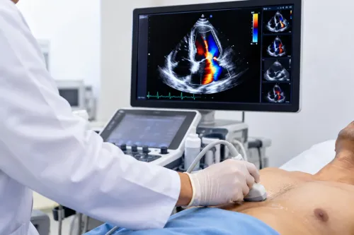
A resting transthoracic 2D echo uses ultrasound to create moving images of the heart without ionising radiation. The sonographer applies gel and moves a transducer across your chest while you lie on your back or left side; the examination usually takes 30 to 60 minutes. So, what does a 2D echo show?
It shows anatomy and movement, not the coronary arteries directly.
A standard study can reveal:
- Chamber size and shape, including enlarged atria or ventricles
- Heart-muscle thickness, which can support assessment of hypertrophy or cardiomyopathy
- Wall motion, including areas that move weakly or do not contract normally
- Ventricular pumping function, commonly reported with the left-ventricular ejection fraction
- Valve structure and function, including narrowing or leakage when Doppler measurements are added
- Congenital structural abnormalities, such as an opening between chambers
- Fluid around the heart, called pericardial effusion
- Selected blood-flow abnormalities, such as an abnormal jet across a valve
A resting echo does not directly display narrowed coronary arteries as coronary CT angiography or invasive angiography does. It can show the effects of heart disease, such as reduced pumping or regional wall-motion changes, but a normal resting result does not answer every question about chest pain.
The label heart ultrasound test in Nashik describes the same underlying technology used elsewhere. Confirm that the booking is for a resting transthoracic echo rather than a stress echo or transoesophageal echo, because those examinations answer different clinical questions.
How Colour Doppler Adds Blood-Flow Information
The 2D image shows the heart’s chambers, walls, valves, surrounding sac and wall motion in real time. It provides the structural picture; Doppler adds information about moving blood.
- M-mode records movement along one narrow ultrasound line over time. It gives precise measurements of chamber dimensions, wall thickness and valve timing, but it samples less area than a 2D image.
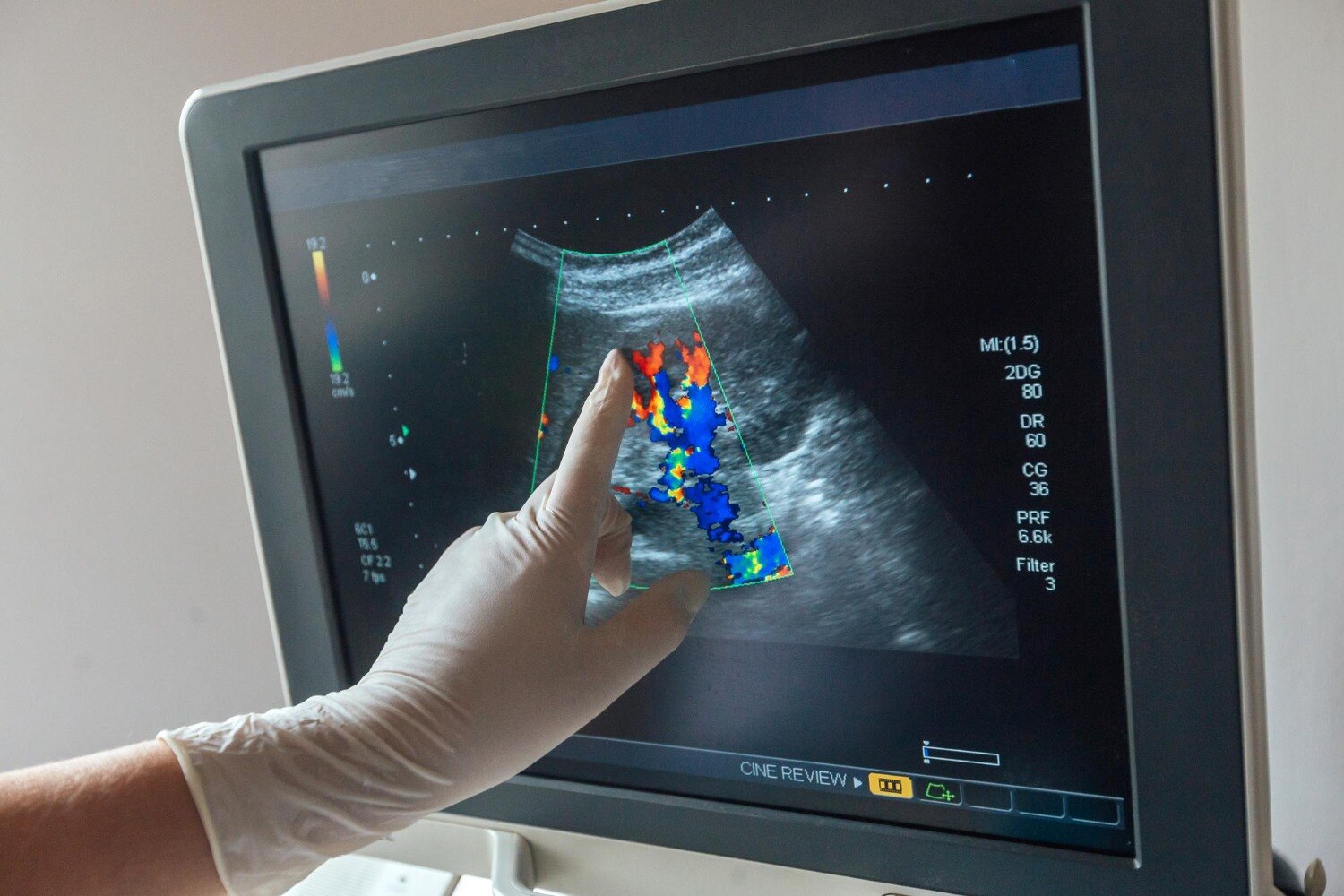
- Colour Doppler overlays moving blood on the heart image. The colours indicate flow direction relative to the probe, not automatically a leak or blockage. Gain, angle, velocity scale and aliasing can change the display.
- Pulsed-wave Doppler measures velocity at a chosen sample location, such as across a valve or within a chamber. It tells the operator where flow occurs, but its measurable velocity range is limited.
- Continuous-wave Doppler measures high velocities along the entire ultrasound beam. It is useful for severe valve narrowing, although it cannot identify the exact point producing the highest velocity.
For echo color doppler test details, remember that colour alone cannot grade severity. The clinician combines the jet’s appearance and width with spectral Doppler, valve anatomy, chamber response and pressure estimates. A gradient is estimated from velocity using ΔP = 4V², so poor beam alignment can significantly distort it.
A search for 2D echo color doppler in Nashik usually refers to this combined examination, not a separate imaging technology.
Which Heart Test Answers Which Question?
Each test answers a different question: a resting 2D echo examines the heart’s structure and mechanical function, while ECG, exercise tests, Holter monitoring and electrophysiology studies focus on electrical activity or performance under stress.
| Test | Main question | What it measures |
|---|---|---|
| Resting 2D echo | What does a 2d echo show? | Chamber size, wall motion, pumping strength, valve structure and function, and fluid around the heart |
| ECG | Is the heart’s electrical activity normal now? | Rhythm, conduction, and patterns suggesting current or previous heart muscle injury |
| TMT | How does the heart’s electrical activity respond to exercise? | Exercise-related ECG changes, symptoms, blood pressure and exercise capacity; it does not create heart images |
| Stress echo | Does exercise or medicine reveal poor blood supply? | Heart-wall motion and valve behaviour during stress; new wall-motion abnormalities can suggest inducible ischaemia |
| Holter monitor | Does an intermittent rhythm problem occur during daily life? | Continuous ECG recording over an extended period, including symptoms and irregular rhythms |
| Electrophysiology study | Where does an abnormal rhythm begin? | Electrical pathways mapped with catheters inside the heart; the team can deliberately trigger arrhythmias |
For 2d echo test details in nashik, confirm whether the booking is for a resting transthoracic echo, colour Doppler, stress echo or transoesophageal echo. A standard transthoracic study usually takes 30 to 60 minutes, uses no ionising radiation and needs no fasting; stress testing has different instructions.
When Echo Is Not the Best Test for the Problem
A resting 2D echo can miss narrowed coronary arteries, early or exertional ischaemia, small areas of scar and rhythm-related problems because it views structure and motion at rest, not the artery lumen or electrical activity. A normal scan does not rule out coronary disease.
Choose the test that matches the unanswered question:
- CT coronary angiography shows the coronary artery walls and lumen, making it suitable for suspected obstructive coronary disease when a non-invasive anatomical view is needed.
- Invasive coronary angiography directly images coronary arteries during catheterisation and allows treatment, such as angioplasty, during the same procedure when appropriate.
- Cardiac MRI gives more detailed tissue characterisation and accurate ventricular volumes. It helps assess scar, inflammation, infiltrative disease and complex cardiomyopathy, but takes longer and may not suit some implanted devices or severe claustrophobia.
- Transoesophageal echo places the probe in the oesophagus, closer to the heart. It provides clearer views of valves, clots, the left atrial appendage and prosthetic valves when chest-wall images are inadequate.
If symptoms occur during exertion, a stress echo can expose new wall-motion abnormalities that a resting scan misses.
Echo color Doppler test details matter: colour alone does not grade a leak or narrowing. The clinician combines jet appearance, spectral Doppler velocity, pressure gradients, flow measurements, chamber response and valve anatomy. Machine gain, settings, insonation angle and aliasing can change the displayed colour.
What to Confirm When Booking a 2D Echo in Nashik
Book the named study, not just a “heart ultrasound test in Nashik.” Ask whether the referral is for a resting transthoracic echocardiogram, a 2D echo with colour Doppler, a stress echo or a transoesophageal echo; each uses ultrasound differently and requires different preparation.
1. Confirm the exact test on the appointment message. “2D echo color Doppler in Nashik” usually means a standard resting echo with colour, pulsed-wave and continuous-wave Doppler, not a separate scan. Stress echo adds exercise or medicine, while transoesophageal echo uses a probe passed into the oesophagus.
2. Ask whether a qualified sonographer will acquire the images and which clinician will interpret the measurements. Request that the report address the referring doctor’s question, such as valve leakage, pumping strength or chamber enlargement.
3. Ask about preparation before you arrive. A routine transthoracic study usually needs no fasting; stress or transoesophageal studies can require fasting, medication instructions or an escort. Follow the facility’s written instructions rather than applying routine 2D echo test details in Nashik to every examination.
4. Confirm the practical timing and clothing. You generally lie on your back or left side while gelled sensors move across your chest. Allow about 30 to 60 minutes; the test uses no ionising radiation.
At Dr Sheetalkumar Hiran at Nine Pearl Hospital, ask the booking desk to record the exact study type and any preparation instructions, rather than relying on the informal word “echo.” Bring previous reports and your referral so the comparison includes earlier measurements.
Related products
 | In 2D Echocardiography, 2D stands for 2 dimensional, Echo means reflection of sound waves cardiography is graph or image of heart and Colour Doppler... View product → |
 | 2-D Echo Color Doppler View product → |
Frequently asked questions
What does a resting 2D echo show?
It shows the heart chambers, walls, valves, pumping movement and some surrounding structures using moving ultrasound images.
What does colour Doppler add to a 2D echo?
Colour Doppler displays blood-flow direction and speed, helping identify valve leakage, narrowing and abnormal flow patterns.
Which heart test should you choose for a specific problem?
An ECG answers questions about electrical rhythm, while a 2D echo assesses structure and movement. Stress testing, Holter monitoring, CT or cardiac MRI address different questions.
When is a 2D echo not the best heart test?
A 2D echo is not the main test for isolated rhythm symptoms, blocked coronary arteries or detailed tissue and vessel imaging. Your clinician may select an ECG, Holter monitor, stress test, CT angiography or cardiac MRI instead.
What should you confirm when booking a 2D echo in Nashik?
Confirm whether the appointment includes colour Doppler, how long the scan takes, whether fasting is needed, when the report will be ready and who will explain the findings.