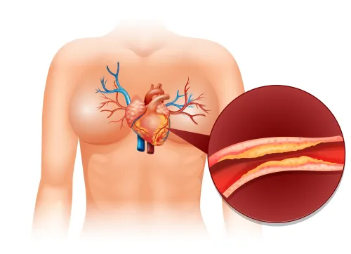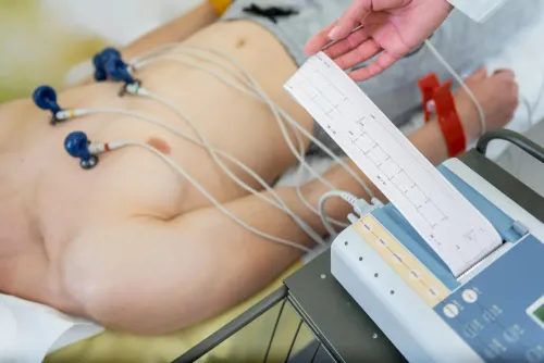


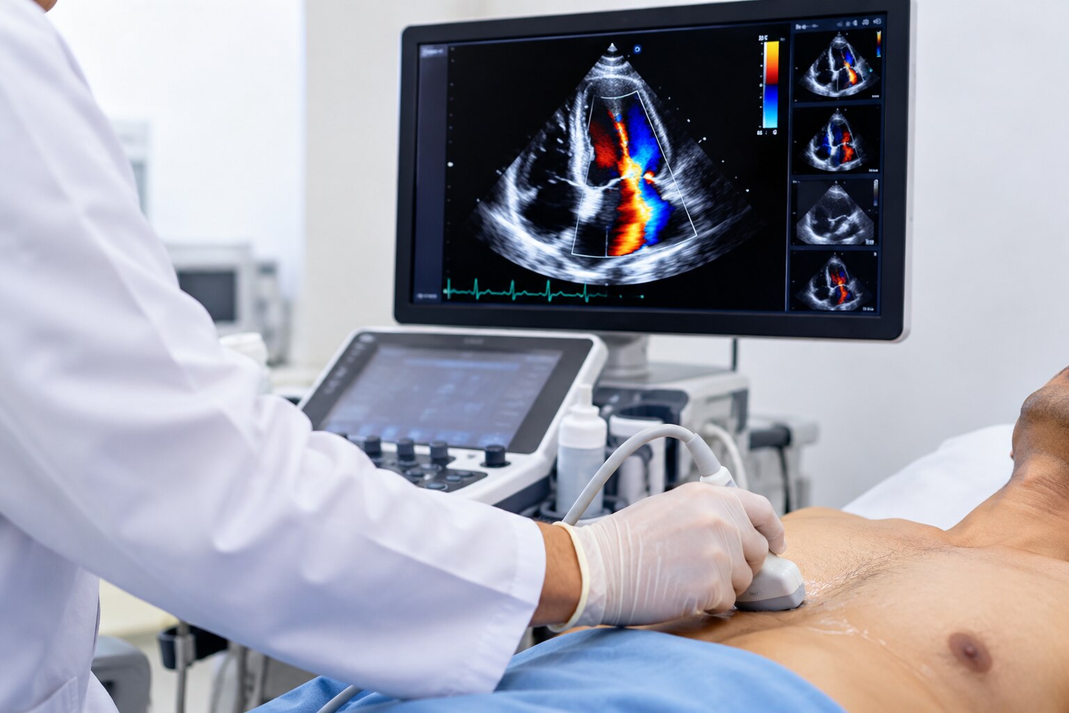
An echo color Doppler checks blood movement inside the heart and across its valves, but it is not a full scan of every artery and vein in the body. By matching your symptoms to the right examination, you can understand whether you need a cardiac study, a vascular Doppler examination, or another form of testing.
Key takeaways
- Echo color Doppler assesses blood flow through heart chambers and valves.
- Vascular Doppler examines blood flow in selected arteries and veins outside the heart.
- Tell the doctor about symptoms, prior heart tests and relevant medical conditions.
- Choose the test based on the suspected blood-flow problem, not the word Doppler alone.
What blood-flow problems can an echo color Doppler find?
An echocardiogram with color Doppler is a non-invasive heart blood flow test. It assesses blood moving through the heart chambers and valves while showing chamber size, wall movement and pumping function. It does not examine every artery or vein in the body.
An echo color Doppler for blood flow problems in Nashik can help identify:
- Valve narrowing, called stenosis, where an opening restricts blood flow.
- Valve leakage, called regurgitation, where blood moves backward.
- An abnormal jet through a hole between heart chambers.
- Some abnormal connections between blood vessels.
The colour display is only one part of the examination. Spectral Doppler records blood-flow velocity over time. Peak velocity and calculated pressure-gradient measurements help assess how severely a narrowed valve obstructs flow.
The study can also estimate pulmonary-artery pressure from the velocity of the tricuspid-regurgitation jet. This is an estimate, not a direct catheter measurement, and accuracy falls when the signal or right-atrial-pressure estimate is unreliable.
A normal resting study does not rule out every cause of chest pain or reduced blood flow to the heart muscle. Depending on your symptoms and examination, a clinician may consider stress imaging, CT coronary angiography or invasive coronary angiography. A report must be interpreted with the clinical picture, not by the colour image alone.
How is a heart Doppler different from a vascular Doppler?
A heart Doppler examines the heart, valves and intracardiac blood flow through the chest. It is an echocardiographic color Doppler and a non-invasive Doppler test for blood flow, not a survey of every vessel in the body.
A vascular Doppler examination uses grayscale imaging, color Doppler and spectral Doppler to assess arteries or veins in the neck, arms, legs or abdomen.
| Test | What it examines | Typical use |
|---|---|---|
| Heart echocardiographic Doppler | Chambers, valves and flow inside the heart | Suspected valve disease or heart-chamber problems |
| Lower-extremity arterial duplex | Leg-artery walls, narrowing, blockage and flow | Cold or painful limb, or peripheral artery disease |
| Venous duplex | Vein compression and altered flow | Leg swelling or suspected deep-vein thrombosis |
For suspected leg-artery disease, an ankle-brachial index is commonly the initial physiologic test. An ABI of 0.90 or less is generally abnormal; calcified, poorly compressible arteries can make the value falsely high. A lower-extremity arterial duplex then helps locate and characterize narrowing or blockage.
For suspected deep-vein thrombosis, venous duplex looks mainly for failure of the vein to compress and altered flow. Vascular velocity measurements depend on a suitable Doppler angle, usually 45 to 60 degrees.
Choose an echo for suspected valve or chamber disease. Choose vascular testing for a cold or painful limb, leg swelling, suspected clot, carotid disease or peripheral artery disease.
When might a doctor order an echo color Doppler?
Breathlessness, chest discomfort, leg swelling, a murmur or fainting can prompt an echocardiogram with Doppler, a non-invasive heart blood flow test. The ordering clinician matches the study to your examination, symptoms and previous results—not to the search term “echo color Doppler in Nashik” alone.
- Breathlessness: check pumping function, chamber size and estimated lung-artery pressure.
- Chest discomfort: assess heart motion and valves; symptoms that appear with exertion may need stress imaging.
- Leg swelling: look for reduced pumping, enlarged chambers or abnormal valve flow.
- Heart murmur: examine valve movement, narrowing and leakage.
- Fainting: assess valve obstruction, pumping function and abnormal intracardiac flow.
- High blood pressure: check chamber enlargement, wall thickness and pressure estimates.
- Suspected valve disease: measure valve movement, leakage and obstruction.
- Known heart disease or treatment follow-up: compare pumping function, chamber size and residual abnormal flow.
| Examination | Main information | When it applies |
|---|---|---|
| 2D echo | Structure and motion | Baseline chamber and pumping assessment |
| Color Doppler | Direction and location of flow | Valve leakage, holes or abnormal connections |
| Spectral Doppler | Flow velocity and pressure-related measurements | Quantifying obstruction or pressure |
| Stress echocardiography | Findings during exercise or medicine | Symptoms absent at rest |
| Transesophageal echo | Closer views through an oesophageal probe | Chest-wall images are inadequate or detail is needed |
The clinician chooses one study or a combination; a resting test is not automatically the right test for exertional symptoms.
What happens during the examination, and how should you prepare?
During an echo color Doppler in Nashik, you usually remove clothing from your upper body and lie on your left side. Gel goes on your chest, and the sonographer moves an ultrasound probe across several chest positions.
1. The sonographer records heart images and Doppler measurements while asking you to turn, hold your breath or breathe in a particular way. Remain still during image capture; the probe can create pressure, but the examination uses no needles and no ionising radiation.
2. Many resting studies do not require fasting. Ask the testing centre whether fasting applies to your appointment, and do not fast beyond its instructions.
3. Wear practical clothing that allows chest access. Ask how long the appointment is expected to take, and confirm whether you should continue routine medicines. Do not stop medicines unless the ordering clinician tells you to.
4. Tell the centre about obesity, lung disease, an unusual chest-wall shape, prior chest surgery or difficulty lying in position. These factors can make ultrasound images technically difficult and may require another approach.
A doppler test for blood flow does not always produce complete images. If the windows are inadequate, the clinician may consider contrast echocardiography, transesophageal echocardiography, CT or MRI rather than treating an incomplete study as normal.
How do you choose the right blood-flow test in Nashik?
Choose the test by locating the suspected problem, not by booking any scan labelled “Doppler.” For breathlessness, a murmur, fainting, swelling with suspected heart disease, valve disease or reduced pumping, ask whether an echocardiographic color Doppler is appropriate.
| Concern | Test to discuss | What it examines |
|---|---|---|
| Heart or valve problem | Echocardiographic color Doppler | Chambers, valves, pumping and flow inside the heart |
| One-sided leg swelling or suspected deep-vein clot | Venous vascular Doppler examination | Vein compression and altered flow |
| Cold or painful limb, reduced arm or leg circulation | Arterial vascular Doppler examination or ABI | Arterial narrowing and circulation |
| Neck-vessel disease | Carotid vascular Doppler examination | Blood flow and narrowing in neck arteries |
A vascular Doppler examination is not the same as an echo color Doppler in Nashik. Doppler velocity results depend on the vessel, ultrasound window, patient position, operator technique and beam angle; vascular measurements usually use a 45-to-60-degree angle. The colour image alone cannot establish severity.
Do not treat a technically limited echo as normal. Obesity, lung disease or poor positioning can block clear views, so the clinician may consider contrast echo, transesophageal echo, CT or MRI.
For an echo color Doppler for blood flow problems in Nashik, confirm availability, preparation rules and report turnaround with the hospital or imaging department. Dr Sheetalkumar Hiran at Nine Pearl Hospital can help direct cardiac assessment, follow-up or referral for another circulation test.
Related products
 | In 2D Echocardiography, 2D stands for 2 dimensional, Echo means reflection of sound waves cardiography is graph or image of heart and Colour Doppler... View product → |
 | 2-D Echo Color Doppler View product → |
Frequently asked questions
What blood-flow problems can an echo color Doppler find?
It can assess blood moving through the heart chambers and valves, including abnormal flow linked to valve problems, and it also shows chamber size, wall movement and pumping function. It does not examine every artery or vein in the body.
How is a heart Doppler different from a vascular Doppler?
A heart Doppler is part of an echocardiogram and evaluates the heart’s chambers, valves and pumping action. A vascular Doppler examination uses ultrasound to assess blood flow in selected arteries or veins elsewhere in the body.
When might a doctor order an echo color Doppler?
A doctor may order it when symptoms, examination findings or another heart test raise questions about valve function, chamber movement, pumping function or blood flow through the heart.
What happens during an echo color Doppler examination?
You lie down while a sonographer places an ultrasound probe on your chest with gel. The examination is non-invasive, and the probe records heart structure, movement and blood-flow patterns.
How do you choose the right blood-flow test in Nashik?
Ask which body area needs assessment and what question the test must answer. Choose an echo color Doppler for suspected heart or valve problems, and a vascular Doppler when the concern involves a named artery or vein.