

Chest pain, an abnormal stress test, or a suspected heart attack does not lead to the same type of angiography or the same timetable. You will learn which symptoms need emergency hospital care, when non-invasive testing comes first, what the procedure involves, and how doctors decide whether its findings will change treatment.
Key takeaways
- Seek emergency care for severe, recurrent chest symptoms or collapse.
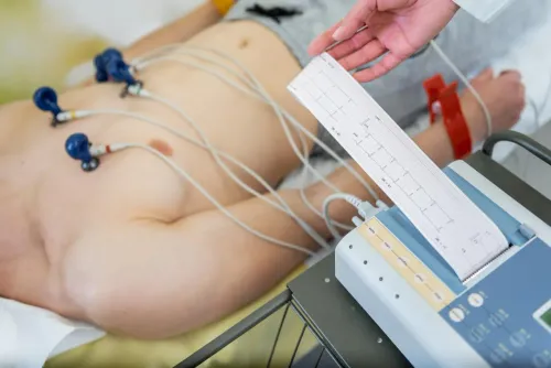
- Troponin, ECG, stress tests, and scans answer different clinical questions.
- CT angiography may assess vessels without catheter insertion.
- An invasive angiogram can lead directly to angioplasty or stenting.
Who needs urgent angiography rather than an outpatient test?
Ongoing or recurrent chest pressure, chest pain, breathlessness, sweating, nausea, fainting, or pain spreading to the arm, jaw, back, or shoulder needs immediate hospital assessment. Do not book an outpatient angiography test in Nashik if symptoms are severe, continue, return repeatedly, or involve collapse.
| Situation | Timing | What usually happens |
|---|---|---|
| Suspected STEMI | Emergency | ECG and clinical assessment can lead to immediate coronary angiography, with angioplasty when available. |
| Non-ST-elevation acute coronary syndrome | Urgent or early, based on risk | Timing depends on very-high-risk features, ECG changes, serial troponin results, ongoing symptoms, ventricular function, and overall clinical risk. |
| Stable suspected coronary disease | Planned | Doctors investigate first and schedule angiography when the result is likely to change treatment. |
A suspected ST-elevation myocardial infarction can require emergency catheter-based angiography to restore blood flow quickly. For non-ST-elevation acute coronary syndrome, everyone does not need an automatic procedure at the same time; very-high-risk findings may require immediate angiography, while others need a risk-based early or selective approach.
Who needs an angiogram on a planned basis? Common reasons include:
- Persistent exertional chest pain despite appropriate medical treatment.
- High-risk or inconclusive stress-test, stress-imaging, or other non-invasive results.
- Known coronary artery disease when doctors need detailed anatomy before angioplasty or bypass surgery.
A normal resting ECG does not rule out coronary disease, and an abnormal test does not automatically mean you need a stent. Low-risk, non-specific symptoms usually call for consultation and non-invasive evaluation before invasive angiography.
How do doctors decide after an ECG, troponin test, TMT, or scan?
A normal resting ECG does not rule out coronary artery disease, and an abnormal stress test does not automatically mean you need a stent. The consultation asks a narrower question: who needs an angiogram, and will its result change treatment?
1. The clinician reviews chest pain pattern, breathlessness, exercise limitation, examination findings, ECG, serial troponin blood tests, and echocardiography. Troponin trends help identify heart-muscle injury; diabetes, smoking, high blood pressure, cholesterol, kidney disease, family history, age, and previous coronary disease alter the likelihood.
2. For stable, low-risk, or non-specific symptoms, coronary CT angiography or stress imaging often comes before an invasive catheter test. These tests provide useful anatomical or blood-flow information without arterial access. The choice depends on age, risk level, ECG findings, exercise capacity, kidney function, contrast concerns, and local expertise.
A TMT is less suitable when you cannot exercise adequately or the ECG makes interpretation unreliable.
3. Invasive angiography becomes more useful when symptoms remain limiting despite medical treatment, testing is strongly abnormal or inconclusive, high-risk anatomy is suspected, or revascularisation is likely. It is also useful when known disease requires anatomical planning before an intervention.
During catheterisation, fractional flow reserve and instantaneous wave-free ratio measure whether an intermediate narrowing actually restricts blood flow. A severe-looking blockage can be functionally unimportant, while symptoms and test results must still be considered together. For angiography for coronary artery disease, the decision is therefore a clinical judgment, not a reaction to one isolated result.
Which imaging test fits a blocked artery outside the heart?
The artery involved determines the imaging choice. An angiography test in Nashik is not automatically a coronary procedure: angiography for vascular disease may examine leg arteries, carotid arteries, kidney arteries, or other peripheral vessels.
| Option | What it shows | When it fits |
|---|---|---|
| Ankle-brachial index and duplex Doppler ultrasound | Pressure differences and blood flow in leg arteries | First assessment for many leg-circulation problems |
| CT angiography | Contrast-enhanced map of arteries | When detailed vessel anatomy is needed quickly |
| MR angiography | Vascular images without ionising radiation | When radiation avoidance matters and device compatibility, availability, and contrast safety are acceptable |
| Catheter-based digital-subtraction angiography | Highly detailed, real-time arterial images | When non-invasive scans are inadequate or an endovascular treatment is planned |
CT coronary angiography is a non-invasive anatomical test of the heart’s arteries. Invasive coronary angiography uses a catheter to show coronary anatomy directly and is more useful when detailed anatomy or treatment planning is needed. A narrowed artery on a scan does not automatically require catheterisation.
For leg symptoms, clinicians often begin with an ankle-brachial index and duplex ultrasound, then use CT angiography or MR angiography if the location or extent of disease remains unclear. Digital-subtraction angiography usually follows only when imaging cannot answer the question or angioplasty, stenting, or another catheter treatment is being considered.
The decision also depends on urgency, kidney function, a previous contrast reaction, implanted devices, and the exact question the doctor needs answered.
What happens before, during, and after an invasive angiogram?
Before arrival for an angiography test in Nashik, the team reviews previous contrast reactions, kidney disease, bleeding problems, anticoagulants, antiplatelet medicines, diabetes medicines, allergies, pregnancy possibility, and fasting instructions. Do not stop prescribed medicines unless the treating team tells you to.
1. On arrival, registration is followed by blood-test review, including kidney function and blood counts. The clinician explains the procedure, alternatives and risks, obtains consent, and connects you to monitoring for blood pressure, heart rhythm and oxygen levels.
2. You lie on an X-ray table. After cleaning the skin, the team injects local anaesthetic, then guides a thin catheter through the radial artery at the wrist or the femoral artery at the groin.
3. Contrast travels through the catheter while X-ray images show the arteries. You may feel brief warmth or flushing. If the findings require angioplasty or stenting, that is a separate treatment decision discussed during the procedure.
4. The team removes the catheter and applies a pressure dressing or compression device. You receive fluids or other hydration advice, remain under observation, and follow instructions about movement, wound care and warning signs. Recovery and discharge timing depend on the access site, findings and whether another procedure was performed.
Risks include bruising or bleeding, arterial injury, contrast reaction and contrast-associated kidney injury. Heart attack, stroke or death are rare. Advanced kidney disease, severe illness or significant vascular disease can increase risk, so the expected benefit must outweigh the procedural risk.
What does an angiogram result mean for your treatment decision in Nashik?
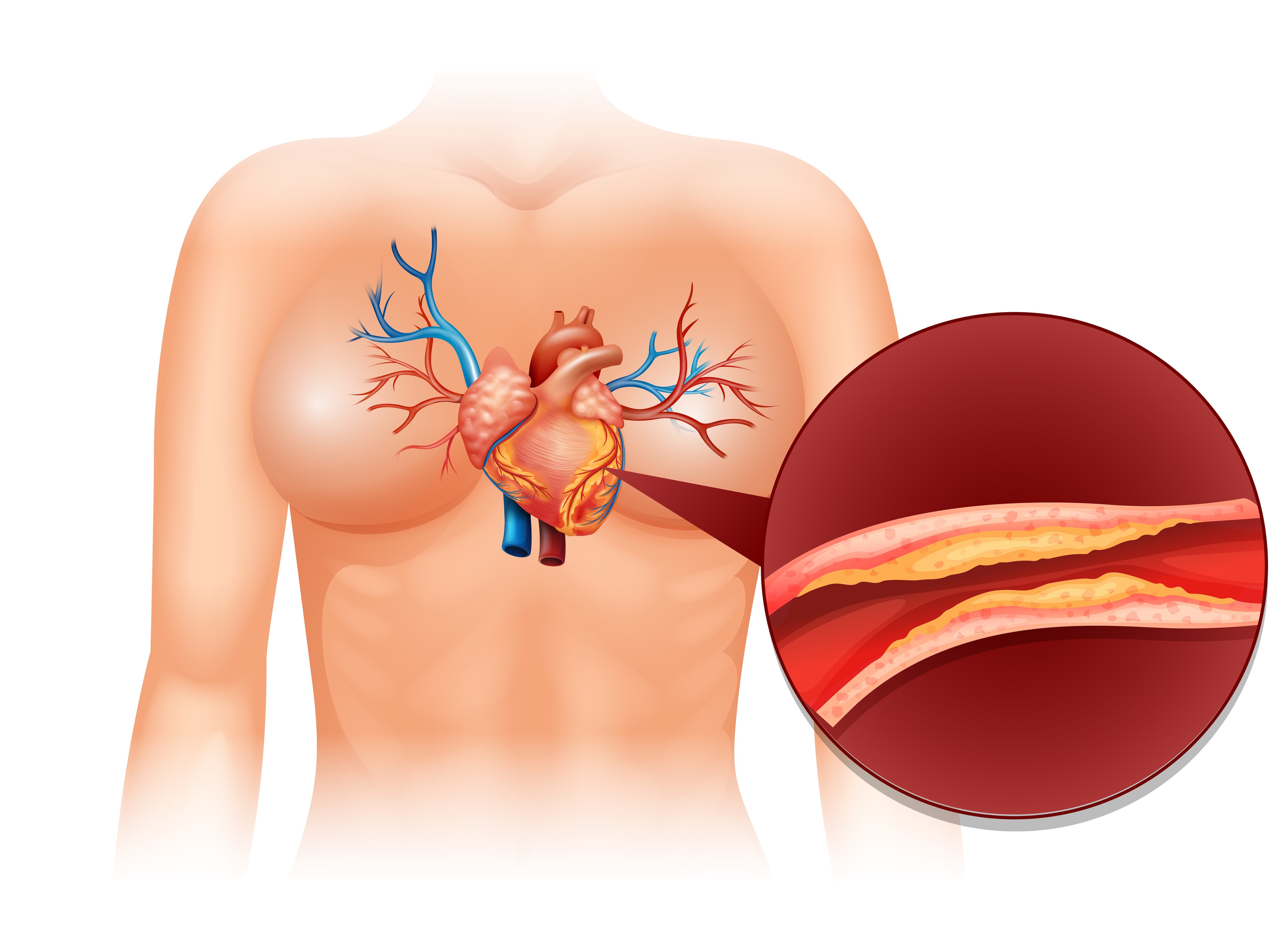
A narrowing on an angiogram does not automatically mean angioplasty or a stent. Angiography for coronary artery disease is diagnostic; the treatment depends on whether the narrowing restricts blood flow and explains the symptoms.
| Option | What it may mean | When it applies |
|---|---|---|
| Medicines and risk-factor treatment | Control cholesterol, blood pressure, diabetes, smoking and symptoms | Stable symptoms or a lesion without significant ischaemia |
| PCI | Balloon treatment with or without a stent | A flow-limiting lesion causing significant symptoms |
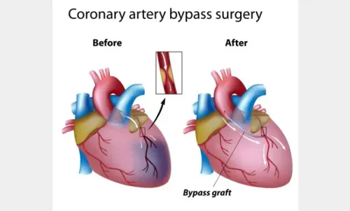
| Bypass surgery | Surgical rerouting around diseased arteries | Selected left-main or multivessel disease, especially with diabetes or reduced left-ventricular function |
| No intervention | Monitoring and investigation of another cause | A functionally insignificant lesion or symptoms not explained by the artery |
A visually tight lesion can still allow adequate blood flow; fractional flow reserve or instantaneous wave-free ratio can clarify an intermediate narrowing. The percentage narrowing alone does not explain every symptom.
A normal coronary angiogram also does not rule out:
- Coronary spasm
- Small-vessel dysfunction
- Reflux
- Lung disease
- Musculoskeletal pain
For angiography for blocked arteries in Nashik, a cardiology consultation at Nine Pearl Hospital with Dr Sheetalkumar Hiran can match urgency, test type, preparation and follow-up to your symptoms and previous results. That coordinated review helps avoid both unnecessary intervention and missed causes of chest pain.
Related product
 | Angiography What is Angiography ? A Coronary Angiogram is a procedure that uses X-ray imaging to see your heart’s blood vessels. |
Frequently asked questions
Who needs urgent angiography rather than an outpatient test?
Seek immediate hospital assessment for ongoing or recurrent chest pressure, chest pain, breathlessness, sweating, nausea, fainting, collapse, or pain spreading to the arm, jaw, back, or shoulder. Do not arrange an outpatient angiography test when symptoms are severe, continue, or return repeatedly.
How do doctors decide after an ECG, troponin test, TMT, or scan?
Doctors combine symptoms, examination findings, ECG changes, troponin results, risk factors, and imaging. ECG and troponin help assess a possible heart attack; TMT evaluates symptoms and blood-flow changes during exertion; scans can show anatomy or reduced blood flow. The combined result determines whether you need urgent invasive angiography, CT angiography, another test, or medical treatment.
Which imaging test fits a blocked artery outside the heart?
The test depends on the vessel and suspected problem. CT angiography can show arteries in the neck, chest, abdomen, pelvis, or legs, while Doppler ultrasound assesses blood flow without radiation. Catheter angiography gives detailed images and allows treatment during the same procedure when intervention is needed.
What happens before, during, and after an invasive angiogram?
Before the procedure, the team checks medicines, allergies, kidney function, pregnancy status when relevant, and fasting instructions. A catheter enters through an artery, contrast is injected, and X-ray images show blood flow. Afterward, staff monitor the puncture site and circulation; follow instructions about fluids, medicines, activity, and warning signs.
What does an angiogram result mean for your treatment decision in Nashik?
A normal result can support medical treatment or investigation of another cause. A narrowing may lead to medicines, lifestyle treatment, monitoring, angioplasty with a stent, or bypass surgery. The decision depends on the artery involved, narrowing location and severity, symptoms, heart function, diabetes, kidney disease, and overall procedural risk.