

A color Doppler echocardiogram can reveal valve narrowing or leakage, abnormal openings between heart chambers, weakened pumping, enlarged chambers and fluid around the heart. You will be able to match symptoms and known conditions to the right type of echo, understand what the test measures, and recognise when emergency care matters more than booking a routine scan.
Key takeaways
- A color Doppler echo assesses valves, chambers, pumping strength and some congenital defects.
- Seek urgent care for severe chest pain, fainting, breathlessness or sudden worsening symptoms.
- Wear comfortable clothing and bring previous heart reports and your current medicine list.
- A resting echo cannot reliably rule out blocked coronary arteries or every rhythm problem.
Which heart problems can a color Doppler echo reveal?
A resting transthoracic color Doppler echo can identify or assess valve disease, abnormal chambers, weak pumping and several congenital defects. If you are asking what can a color Doppler echo detect in Nashik, the answer depends on the images obtained and the clinical question.
- Aortic or mitral stenosis, including restricted valve opening and raised pressure gradients
- Mitral, aortic or tricuspid regurgitation, shown by backward leakage and altered blood-flow direction and velocity
- Prosthetic-valve dysfunction and infective endocarditis-related valve damage
- Enlarged chambers, cardiomyopathy, reduced pumping function and regional wall-motion abnormalities
- Atrial septal defects, ventricular septal defects and patent ductus arteriosus, which create abnormal shunts
- Pericardial effusion and possible tamponade signs, such as right-sided chamber compression and abnormal breathing-related flow changes
- Clues suggesting pulmonary hypertension, including right-ventricular enlargement or dysfunction
The color Doppler heart test measures left-ventricular ejection fraction, chamber dimensions, wall motion, valve opening and leakage, pressure gradients, shunts, and blood-flow direction and velocity. A normal ejection fraction does not exclude heart failure with preserved ejection fraction.
| Finding | What it means | Important limit |
|---|---|---|
| Pulmonary pressure | Estimated from the tricuspid-regurgitation jet | It is not a direct pressure measurement; right-heart catheterisation confirms pressure when necessary |
| Aortic root | Visible enlargement can be assessed | A routine study cannot reliably screen the entire aorta |
| Color jet | Shows flow direction and leakage | Severity requires Doppler measurements, valve anatomy and chamber response, not colour alone |
Who needs an echocardiogram, and when is it urgent?
A person with a new or unexplained heart murmur, breathlessness, leg swelling, fainting, or an abnormal ECG may need an echocardiogram.
Other indications include chest pain with suspected structural disease, high blood pressure with suspected heart effects, known valve disease, heart-failure symptoms, suspected cardiomyopathy, congenital heart disease, or follow-up after a cardiac event, procedure, or treatment.
1. Request an echo test for heart problems when the clinician needs to answer a defined question, such as whether a valve leaks, pumping is reduced, or fluid surrounds the heart. Do not repeat the scan automatically at fixed intervals without a reason; the timing depends on symptoms, examination findings, and the condition being monitored.
2. Treat severe or persistent chest pressure, sudden breathlessness, fainting, blue lips, or new one-sided weakness as emergency symptoms. Seek emergency assessment rather than booking a routine echo appointment, because the scan alone cannot provide the immediate treatment these conditions may require.
3. An echo finding suggestive of cardiac tamponade, such as right-sided chamber compression, also needs immediate clinical correlation. Clinicians must check blood pressure, pulse, symptoms, and the bedside examination because falling circulation can make tamponade life-threatening.
A stable murmur, gradually worsening breathlessness, or planned valve follow-up can follow routine referral, but worsening symptoms should change that plan.
What happens during the test, and how should you prepare in Nashik?
Most standard transthoracic scans take about 30 to 60 minutes. They are painless, although pressure from the probe can feel firm.
1. Change from the waist up and lie on your left side. This position brings the heart closer to the chest wall.
2. The sonographer attaches small electrodes to your chest to monitor the heartbeat during imaging.
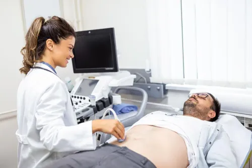
3. Gel is applied to your chest. The sonographer moves a handheld probe between your ribs, changing its position and angle while applying pressure.
4. The probe captures moving heart images and Doppler recordings of blood-flow direction and speed. You may need to hold your breath briefly or shift position.
Fasting, regular medicines, clothing and arrival instructions depend on the exact test and local protocol. Confirm these details when scheduling 2d echo color doppler in nashik, especially for an appointment with Dr Sheetalkumar Hiran at Nine Pearl Hospital; do not stop prescribed medicines unless the facility tells you to.
A routine transthoracic study usually needs no sedation. A transesophageal echocardiogram is different: its probe passes into the oesophagus, so it requires throat anaesthesia, sedation planning, an escort home and a hospital or procedure setting. Get those instructions directly from the facility before attending.
Related products
 | 2D Echo Color Droppler In 2D Echocardiography, 2D stands for 2 dimensional, Echo means reflection of sound waves cardiography is graph or image of heart and Colour Doppler... |
 | 2-D Echo Color Doppler 2-D Echo Color Doppler |
Which type of echocardiogram answers your question?
A resting transthoracic echocardiogram (TTE) is the usual chest-wall scan for chambers, valves, pumping strength and blood flow. It is the examination most people mean by a 2D echo color Doppler in Nashik, but it is not the right answer to every echo test for heart problems.
| Test type | What it examines | When it is chosen |
|---|---|---|
| Resting transthoracic echo | Moving images of chambers, valves, heart muscle and Doppler flow through the chest wall | Suspected valve disease, weak pumping, enlarged chambers or structural defects |
| Transesophageal echo | Closer views from a probe placed in the oesophagus | When clearer images are needed for valves, clots, prosthetic valves, septal defects or parts of the aorta |
| Stress echocardiogram | Heart function during exercise or medicine-induced stress | To look for stress-related wall-motion changes; it is not the same as a resting study |
| Fetal echocardiogram | A developing baby’s heart during pregnancy | When screening, family history or pregnancy factors raise concern |
Your referral should name both the clinical question and the test type. “Assess valve leakage with a resting TTE” is more useful than “echo,” because the wrong examination can leave the question unanswered.
Ask whether the appointment includes contrast, a bubble study or sedation. Contrast can improve weak images, while a bubble study looks for certain abnormal passages of blood. Sedation planning applies particularly to transesophageal echocardiography, not a routine chest-wall scan.
What can a routine resting echo miss?
A routine resting transthoracic color Doppler echo is a snapshot of heart structure and function, not a complete heart check. It does not reliably diagnose most coronary artery blockages, intermittent rhythm disorders, early coronary disease, every cause of chest pain or all myocarditis.
A normal ejection fraction also does not rule out heart failure with preserved ejection fraction (HFpEF).
A small atrial or ventricular septal defect can be missed or underestimated. If suspicion remains, a bubble study, transesophageal echo or cardiac MRI may be needed.
Color jets alone cannot grade a leak: gain settings, aliasing, eccentric jets and loading conditions change how regurgitation appears, so clinicians combine quantitative Doppler measurements with valve anatomy and the heart chambers’ response.
Poor images from obesity, chronic lung disease or positioning problems can make a color Doppler heart test technically limited. Contrast echo, transesophageal echo, cardiac MRI or CT may provide better evidence; an incomplete scan should not be treated as a normal result.
The next test depends on the question:
- CT coronary angiography looks for coronary narrowing; invasive angiography gives catheter-based coronary detail.
- ECG monitoring looks for intermittent rhythm disorders.
- Blood tests help assess injury, inflammation or causes of breathlessness.
- Stress imaging looks for function changes triggered by exertion or medication.
An echo test for heart problems is valuable, but its limits belong in the interpretation.
Frequently asked questions
What heart problems can a color Doppler echo detect?
It can identify or assess valve narrowing and leakage, enlarged or abnormal chambers, reduced pumping function, selected congenital defects and abnormal blood flow between chambers.
Who needs an echocardiogram, and when is it urgent?
You may need one for breathlessness, chest pain, swelling, a heart murmur, fainting, suspected valve disease or known heart disease. Seek urgent medical care for severe chest pain, sudden breathlessness, fainting or rapidly worsening symptoms.
What happens during a color Doppler echo?
You lie on your side while a technician places an ultrasound probe with gel on your chest. The test is non-invasive and usually requires no fasting; bring prior reports and your medicine list.
Which type of echocardiogram answers your question?
A resting transthoracic echo is the usual starting test. A stress echo assesses the heart during exertion, while a transoesophageal echo provides closer views when chest images are inadequate or detailed valve assessment is needed.
What can a routine resting echo miss?
It may not show coronary artery blockages directly, intermittent rhythm disturbances or problems that appear only during exercise. Your clinician may recommend an ECG, stress test, CT coronary angiography or another investigation.